Visualization of a Limonene Synthesis Metabolon Inside Living Bacteria by Hyperspectral SRS Microscopy
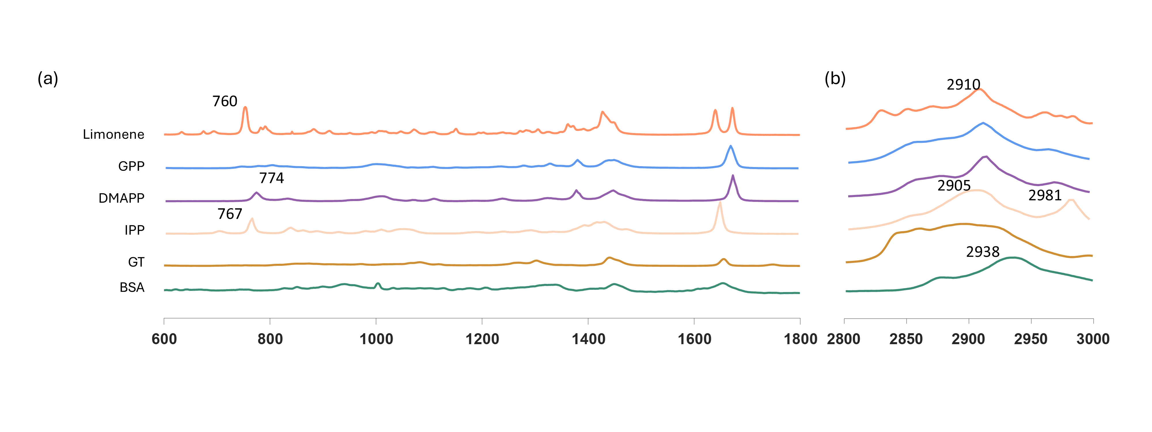
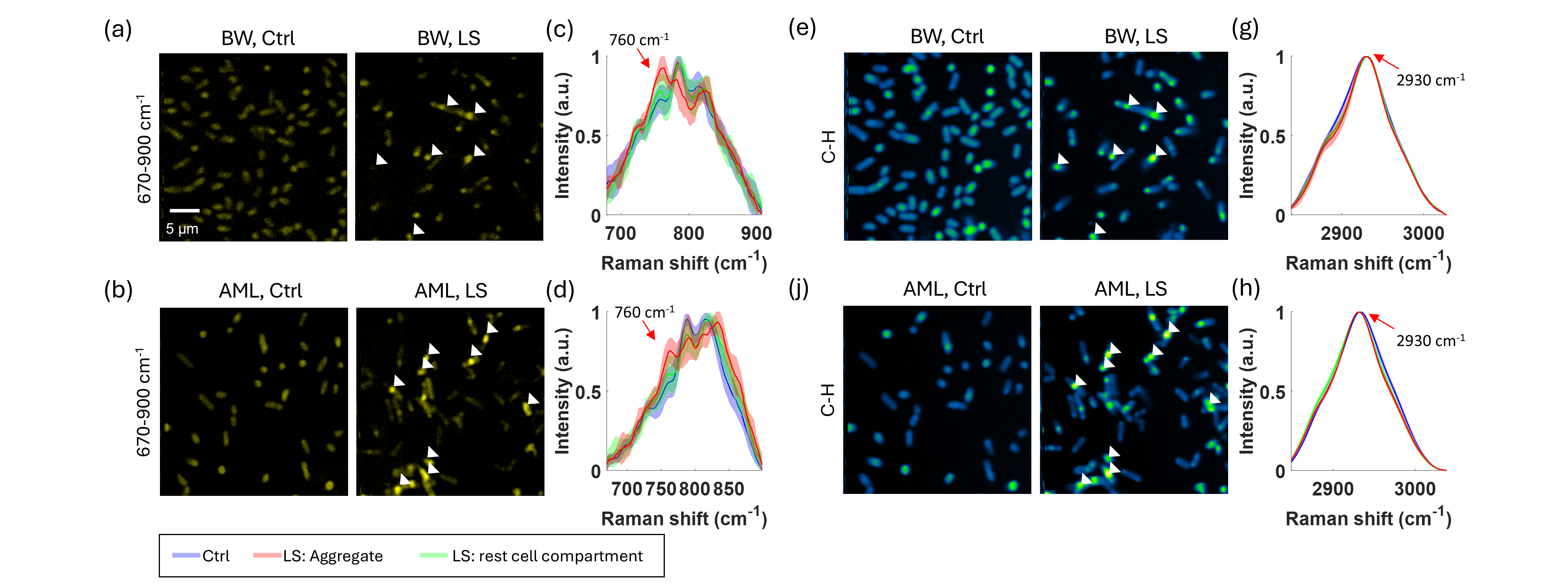
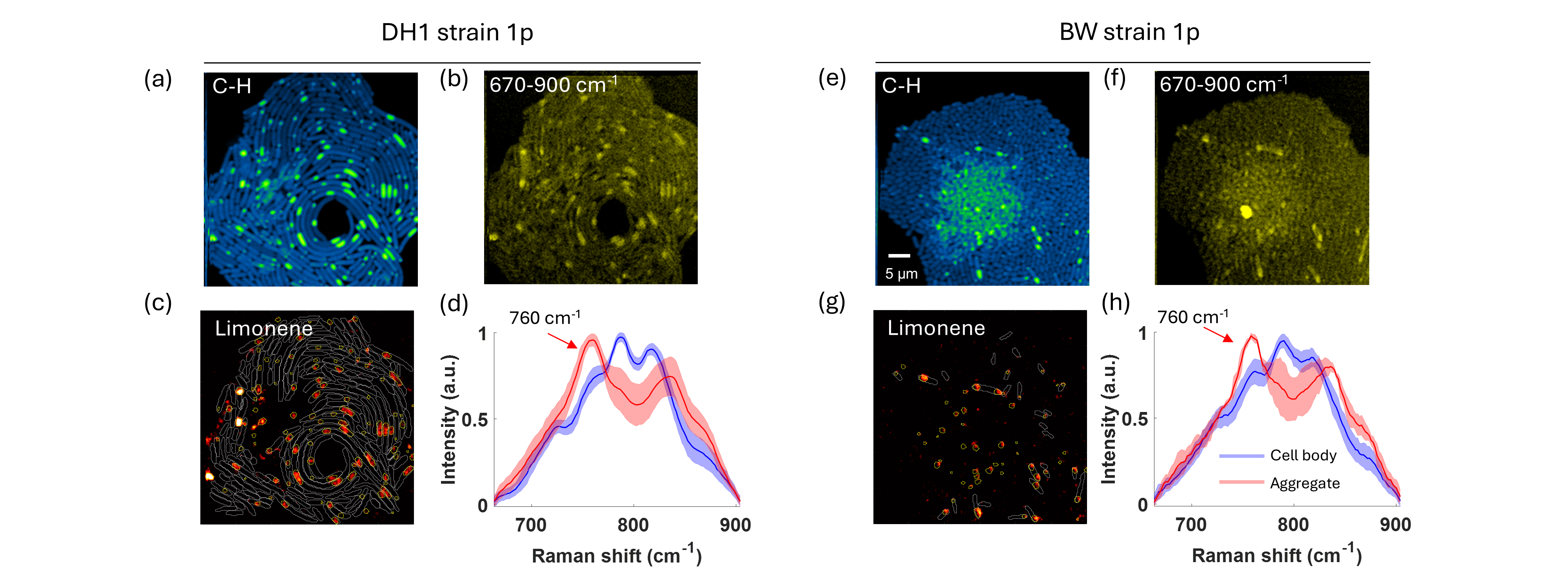
Monitoring biosynthesis activity at single-cell level is key to metabolic engineering but is still difficult to achieve in a label-free manner. Using hyperspectral stimulated Raman scattering imaging in the 670–900 cm−1 region, localized limonene synthesis are visualized inside engineered Escherichia coli. The colocalization of limonene and GFP-fused limonene synthase is confirmed by co-registered stimulated Raman scattering and two-photon fluorescence images. The finding suggests a limonene synthesis metabolon with a polar distribution inside the cells. This finding expands the knowledge of de novo limonene biosynthesis in engineered bacteria and highlights the potential of SRS chemical imaging in metabolic engineering research.
Our paper can be found at Adv. Sci. 2022
We also presented the study at BMES2022 Conference.